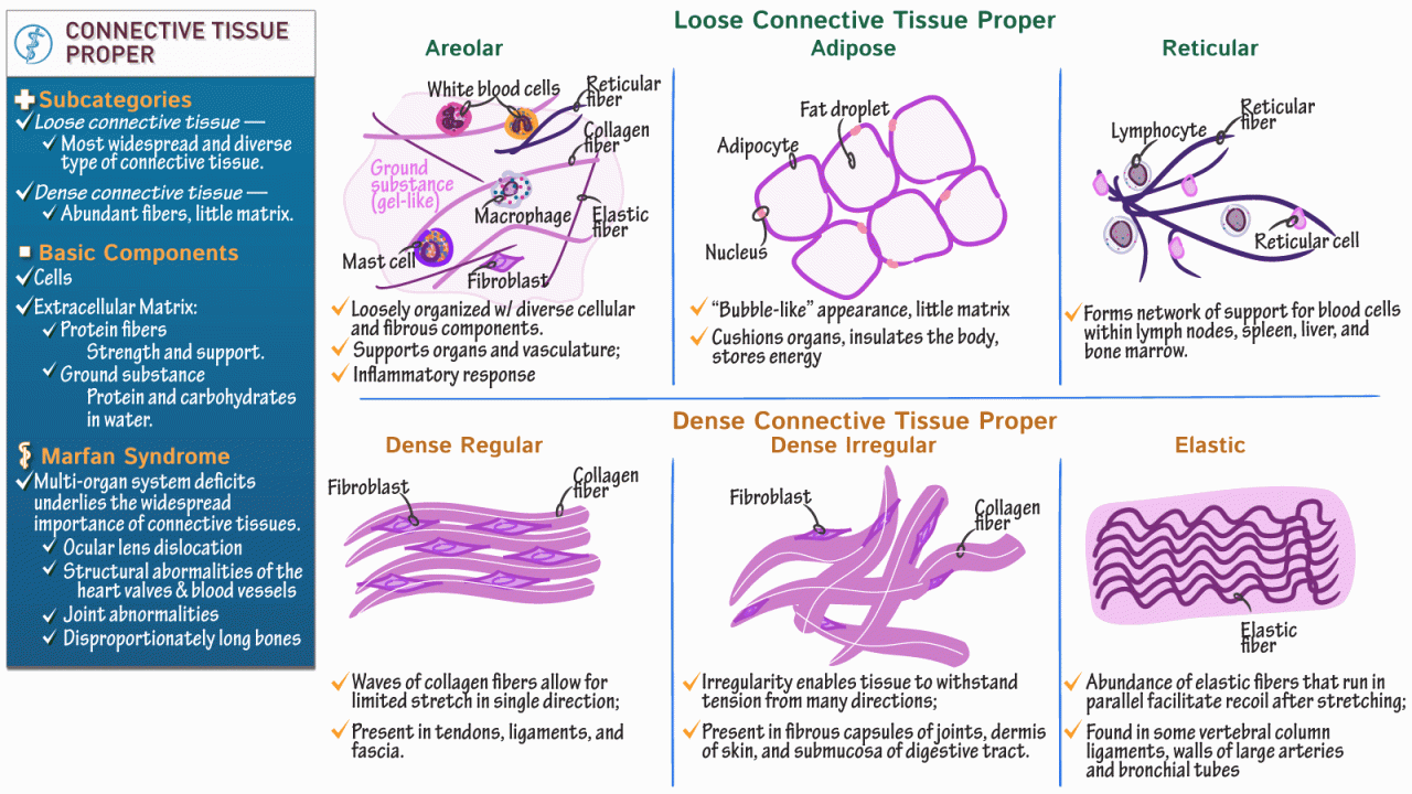
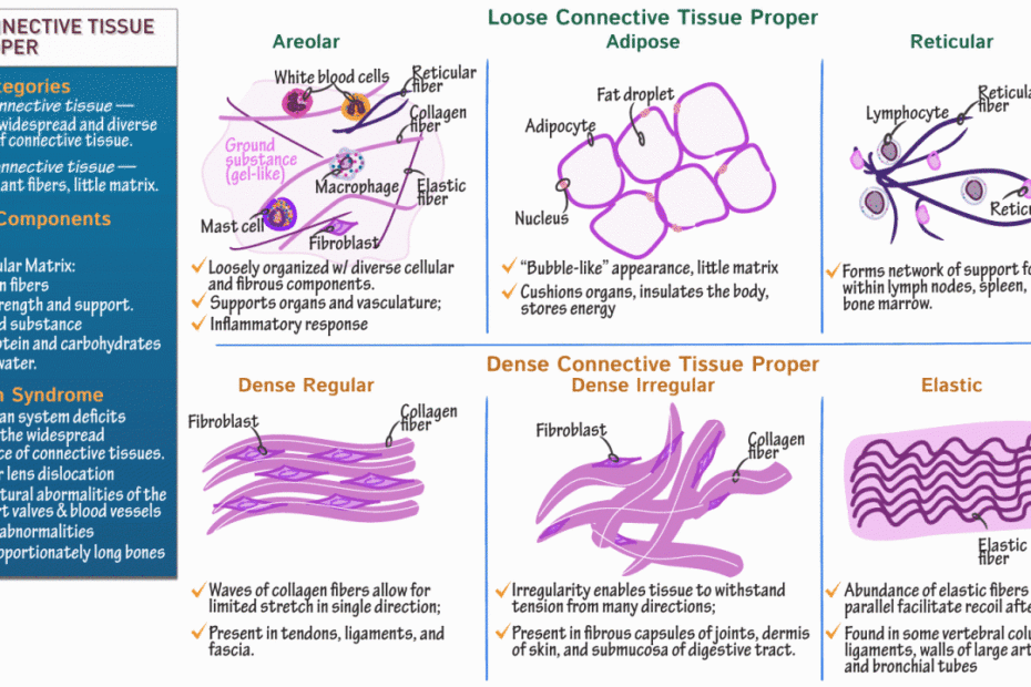
In this topic, we will explore the various aspects of reticular connective tissue drawing, including its structure, function, and importance. We will delve into the finer details of how this complex tissue is formed and examine its role in supporting the various organs and structures in which it is found. Additionally, we will discuss the techniques used to draw reticular connective tissue accurately and effectively, including the use of various types of microscopy and staining methods.
Overall, the study of reticular connective tissue drawing is an important area of study in anatomy and histology that provides a deeper understanding of the complex workings of the human body. By exploring this topic, we can gain valuable insights into the vital functions of our organs and structures and the role of reticular connective tissue in maintaining their health and integrity.
Found 47 images related to reticular connective tissue drawing theme


:max_bytes(150000):strip_icc()/dense_connective_tissue-56a09aee3df78cafdaa32ca1.jpg)


reticular connective tissue drawing
The human body is made up of different types of connective tissues that serve various purposes, such as supporting, connecting, and protecting different organs and tissues. One of these connective tissues is the reticular connective tissue, which is characterized by the presence of reticular fibers and cells.
Types of Reticular Connective Tissue
There are two types of reticular connective tissue, namely the loose reticular connective tissue and the dense reticular connective tissue. The loose reticular connective tissue is composed of a loose network of reticular fibers and cells, while the dense reticular connective tissue has a more compact network of reticular fibers.
Components of Reticular Fibers
Reticular fibers are thin fibers that are made up of collagen protein. They are produced by specialized cells called reticular cells, which are located within the reticular connective tissue. The reticular cells produce collagen protein and secrete it into the extracellular matrix, where it forms the reticular fibers.
Location of Reticular Connective Tissue in the Human Body
Reticular connective tissue is found in various parts of the human body, including the liver, spleen, lymph nodes, bone marrow, and adipose tissue. In the liver, reticular connective tissue forms a network of fibers that support the hepatic cells and regulate blood flow. In the spleen, it forms a network of fibers that support the circulation of blood and the function of the immune system. In the bone marrow, it forms a network of fibers that support the differentiation and maturation of blood cells.
Functions of Reticular Connective Tissue
The main function of reticular connective tissue is to provide support and structure to different organs and tissues in the body. The reticular fibers and cells form a scaffolding that supports the growth and development of various tissues such as the red and white blood cells in the bone marrow. The reticular fibers also aid in the formation of the extracellular matrix, which provides a framework for the other tissues and organs in the body.
Clinical Significance of Reticular Connective Tissue
Reticular connective tissue is associated with several diseases and conditions, including fibrosis, lymphedema, and cancer. Fibrosis is a condition where the excess deposition of collagen fibers in the reticular connective tissue leads to the destruction of organs and tissues. Lymphedema is a condition where the accumulation of fluid in the lymphatic system causes swelling and inflammation in the tissues. Cancer cells can also invade the reticular connective tissue, leading to the growth and metastasis of tumors.
FAQs
1. What is hyaline cartilage, and how is it different from reticular connective tissue?
Hyaline cartilage is a type of connective tissue that is composed of chondrocytes and extracellular matrix. It is found in various parts of the body, including the joints, ribs, and nasal septum. Hyaline cartilage has a rubbery texture and provides cushioning and support to the joints and other structures. Reticular connective tissue, on the other hand, is characterized by the presence of reticular fibers and cells, which provide support and structure to various organs and tissues, including the liver, spleen, and lymph nodes.
2. What is dense regular connective tissue, and how is it different from dense irregular connective tissue?
Dense regular connective tissue is a type of connective tissue that is composed of collagen fibers that are arranged parallel to each other. It is found in structures such as tendons and ligaments and provides tensile strength and flexibility to these structures. Dense irregular connective tissue, on the other hand, is composed of collagen fibers that are arranged in a random, disorganized fashion. It is found in structures such as the dermis of the skin and provides strength and support to these structures.
3. Can you label the different components of connective tissue in a drawing?
Yes, a connective tissue drawing can be labeled to show the different components, including the extracellular matrix, collagen fibers, elastic fibers, and ground substance. Additionally, specific types of connective tissues, such as reticular connective tissue, bone connective tissue, and adipose connective tissue, can be labeled to show their unique features and functions.
4. What is the function of bone connective tissue, and how is it different from reticular connective tissue?
Bone connective tissue is composed of osteocytes, osteoblasts, and osteoclasts, and it provides support and structure to the skeletal system. The extracellular matrix of bone connective tissue is made up of collagen fibers and mineral salts, which provide strength and rigidity to the bone. Reticular connective tissue, on the other hand, provides support and structure to various organs and tissues, including the liver, spleen, and lymph nodes. The reticular fibers and cells of the reticular connective tissue form a network that supports the growth and development of these tissues.
5. What is adipose connective tissue, and how is it different from reticular connective tissue?
Adipose connective tissue is a specialized type of connective tissue that is composed of adipocytes, which store energy in the form of fat. Adipose tissue provides thermal insulation, cushioning, and energy storage to the body. Reticular connective tissue, on the other hand, is characterized by the presence of reticular fibers and cells, which provide support and structure to various organs and tissues, including the liver, spleen, and lymph nodes. The reticular fibers and cells of the reticular connective tissue form a network that supports the growth and development of these tissues.
Keywords searched by users: reticular connective tissue drawing hyaline cartilage drawing, dense regular connective tissue drawing, dense irregular connective tissue, connective tissue drawing with label, reticular connective tissue function, bone connective tissue drawing, adipose connective tissue, reticular tissue
Tag: Top 30 – reticular connective tissue drawing
DRAW -Reticular Connective tissue -Dense Irregular CT, Dense Rregular CT (Tendon + Ligament)
See more here: khoaluantotnghiep.net
Article link: reticular connective tissue drawing.
Learn more about the topic reticular connective tissue drawing.
- Reticular Tissue – Spleen | Math, Tissue, Drawings
- Reticular connective tissue – Histology – Kenhub
- Reticular Connective Tissue – Histology Laboratory Manual
- Reticular Connective Tissue – an overview – ScienceDirect.com
- Connective tissues – Histology – Mavs Open Press
- Reticular connective tissue 1. Reticular fibers. 2. …
- Reticular connective tissue – Histology – Kenhub
- Reticular connective tissue
- Reticular connective tissue – Wikipedia
- 238 Reticular Tissue Images, Stock Photos & Vectors
- Reticular Connective Tissue – ScienceDirect.com
Categories: khoaluantotnghiep.net/wikiimg
